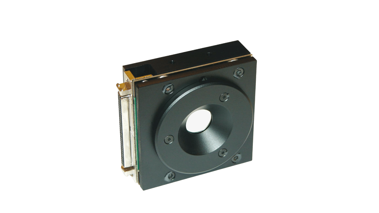
Mirao 52e deformable mirrors
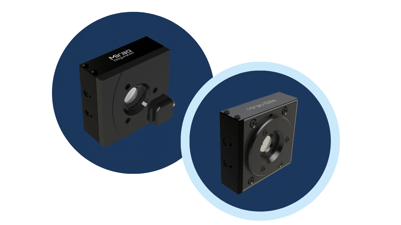
3 versions for various applications
Description
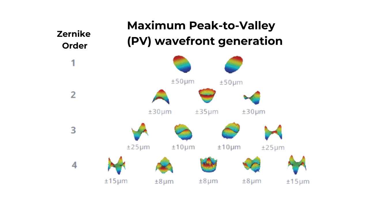
It also provides a 50μm PV deformation amplitude, making it a useful tool for various bio-imaging, microscopy and ophthalmology applications where you will need to correct large-amplitude aberrations.
For use over a long period of time, a high stabilization option is available : Mirao regulated.
You can also ask for a dust proof version : Mirao protected
Technology & features
Principle & implementation
Electronic unit connection to the PC via USB port
Trigger output available to facilitate synchronization with other devices via TTL signal
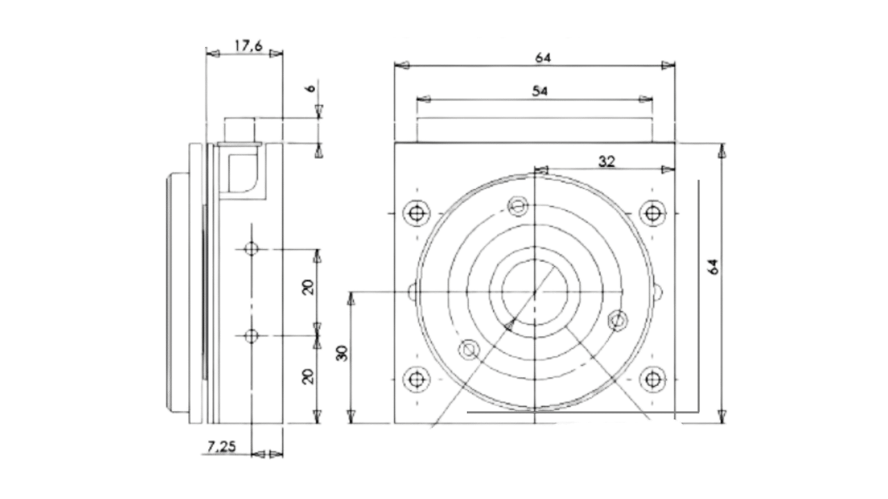
Advantages of Mirao 52e
Closed-loop precision reaching 7nm RMS
Virtually no hysteresis (less than 2%)
Nearly perfect linearity (more than 95%)
Dust proof & high stabilization options available
About the software
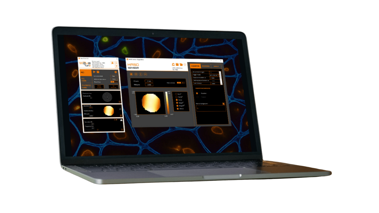
For easy and fast implementation, we recommend using WaveTune, program controls all the elements with a simple user interface
For implementation of aberration detection methods into home-built software, we also provide WaveKit Bio, the Software Development Kit (SDK)
Gallery
brain: GCaMP7-labeled neurons involved in the circadian clock networkimaged at 40µm depth. AO enables the visualization of neuronal projections (white arrows). Courtesy of A. Hubert (École Supérieure de Physique et de Chimie Industrielles, Imagine Optic)
Selected publications
Webinars & videos
Related videos coming soon


