MicAO, an adaptive optics module
for super resolution microscopy
Increase resolution
by correcting aberrations
Shape your PSF
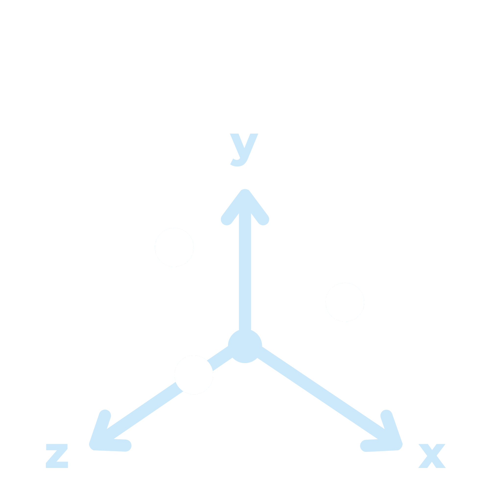
and go 3D

Image deeper
up to 50µm depth
Description
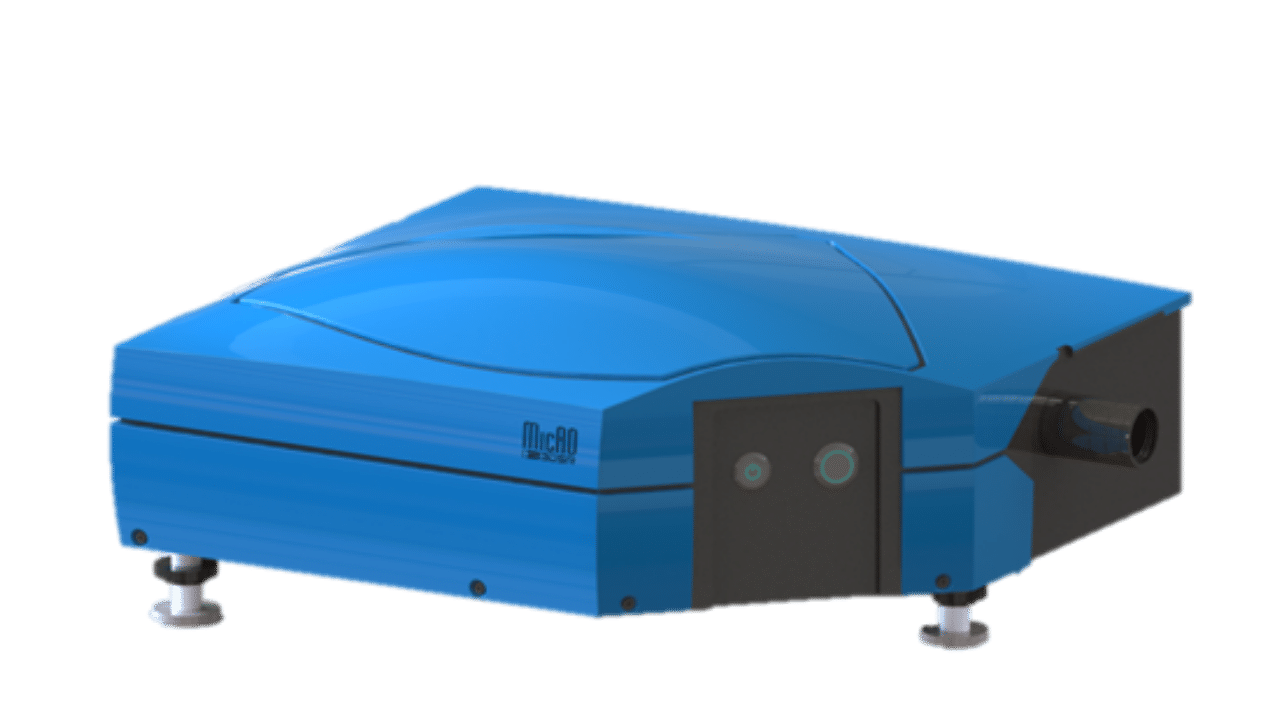
MicAO Adaptive Optics (AO) device is the first AO add-on module dedicated to techniques such as PhotoActivation Localization Microscopy (PALM), Stochastic Optical Reconstruction Microscopy (STORM) and Single Particle Tracking (SPT).
This module will enable you to get a better resolution in both 2D&3D for Single Molecule Localization Microscopy (SMLM).
Technology & features
How is it implemented ?
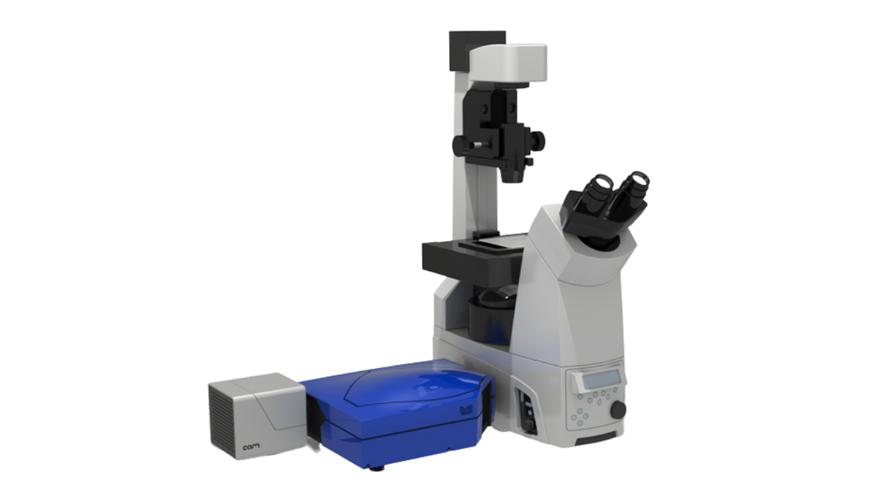
Added between the side port of your inverted frame microscope and the imaging camera
Functions as an image relay : allows you to add other modules (dual color view device, microscope port splitters…)
Compatible with 60x and 100x objective lenses
Advantages of MicAO
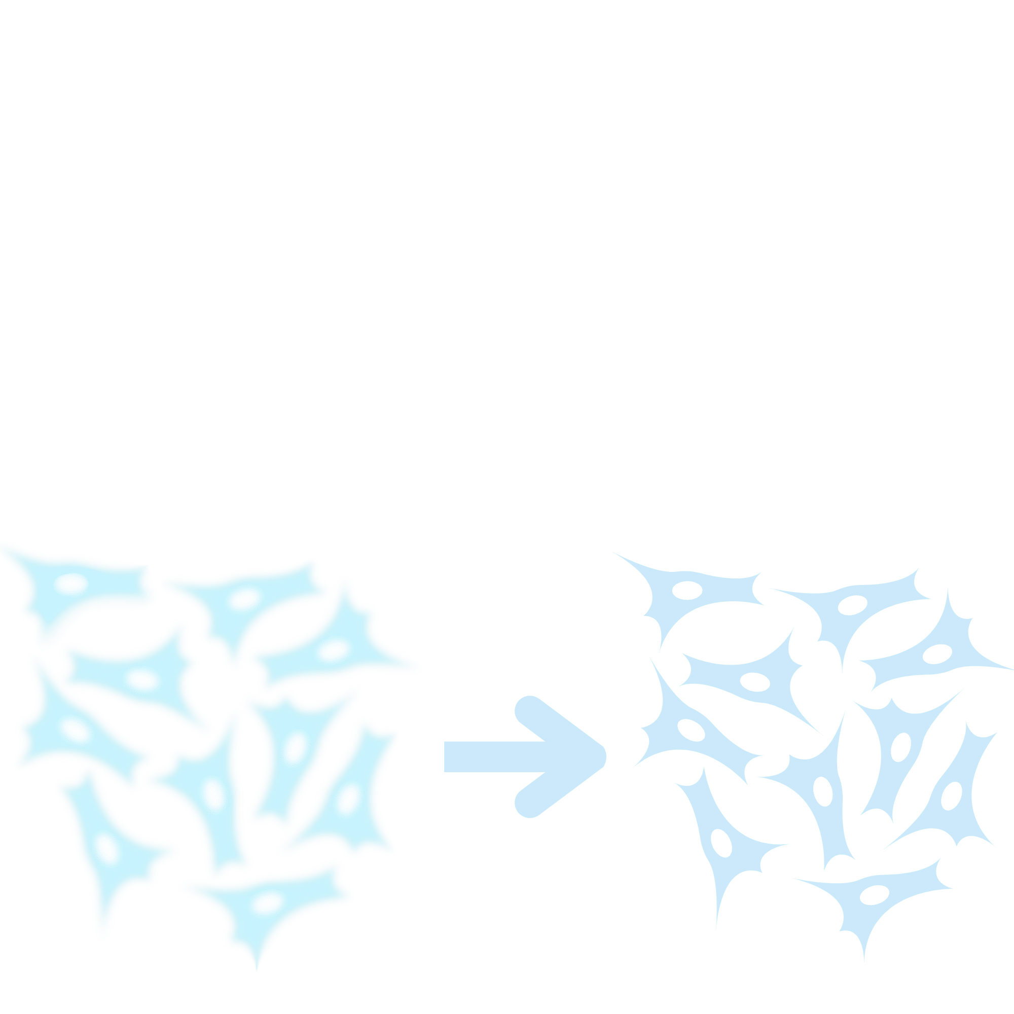
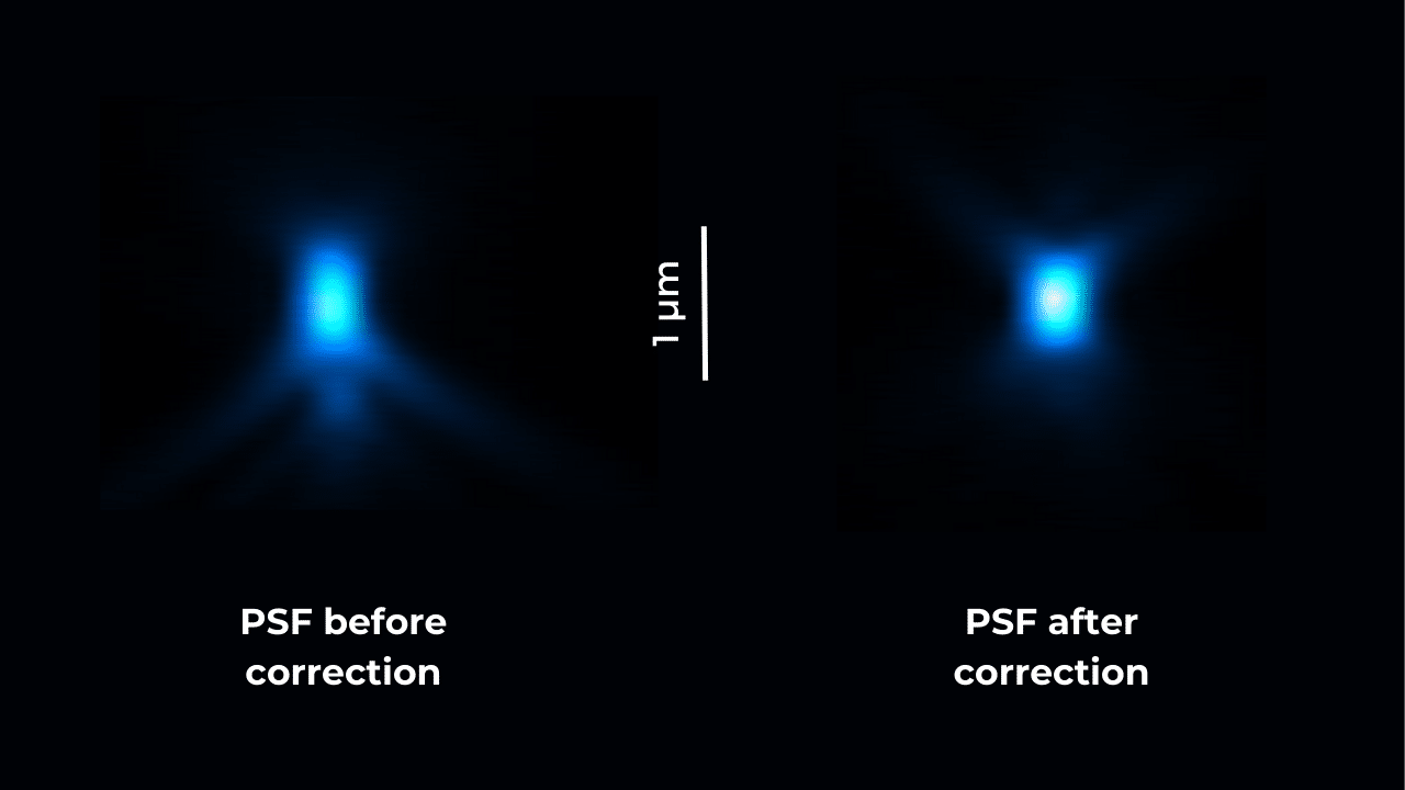
Restores Point Spread Function (PSF) symmetry
Doubles the number of detected photons
Improves near diffraction-limited resolution
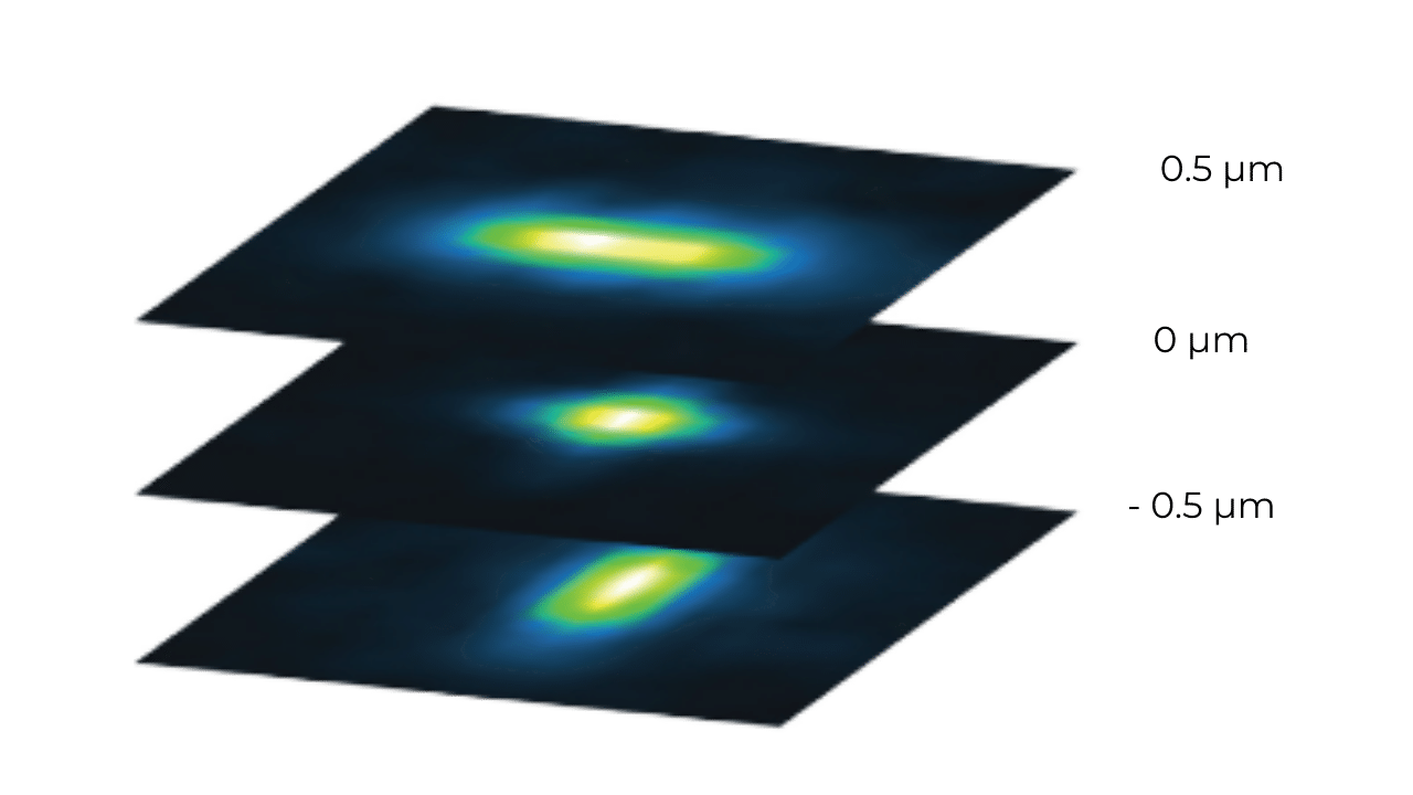
Allows you to create a perfect PSF ecoding astigmatism or tetrapod to go 3D
Gives you a better 2D and 3D localization precision
Is compatible with long-term imaging
How does it work ?
Enables you to function in open-loop configuration
Helps you to apply 3N alogorithm to detect and correct aberrations, thanks to MicAO software
Allows you to control your mirror and shape your PSF
Gallery
Selected publications
Webinars & videos
WEBINAR – PSF Optimization for 3D Super Resolution Microscopy
WEBINAR – Adaptive Optics in SMLM for deep tissue imaging