Increasing the speed of sensorless Adaptive Optics: phase diversity applied to fluorescence microscopy
published on 05.19.2025

Sensorless Adaptive Optics (AO) for fluorescence microscopy
In the last decade, multiple AO methods have been demonstrated in fluorescence microscopy to counteract optical aberrations of biological tissue and restore good image quality, especially in depth. Among these methods, sensorless AO is often used, due to its easier implementation and lower instrumental complexity.
Indeed, the method only requires the integration of a wavefront modulator (no need for a wavefront sensor) and the use of widely available iterative algorithms.
However, this apparent simplicity comes at the cost of several limitations, the most annoying one being the need for tens of images during several algorithm iterations to provide a correction in most implementations, usually corresponding to tens of seconds. This additional time requirement severely increases phototoxicity, and limits fast (3D) imaging.
Phase diversity only requires a few images
In response to this limitation, a publication in Optica (Courtney Johnson, Min Guo, Magdalena C. Schneider, Yijun Su, Satya Khuon, Nikolaj Reiser, Yicong Wu, Patrick La Riviere, and Hari Shroff, “Phase-diversity-based wavefront sensing for fluorescence microscopy,” Optica 11, 806-820 (2024)) from H. Shroff and collaborators proposed an alternative sensorless AO approach, based on Phase Diversity (PD). Specifically, researchers tailored previous PD approaches originally mainly developed for astronomy to the requirements and constraints of fluorescence microscopy.
Essentially, based on a model of image formation in microscopy, the approach introduces known amounts of selected aberrations to generate additional images of the sample, and then uses these images as an input to a Gaussian-Newton optimization process to find the wavefront that minimizes a least-squares objective function.
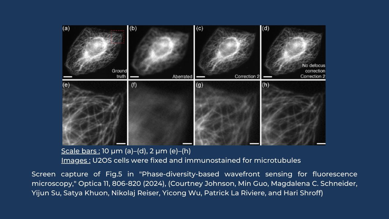
Efficient aberration correction leading to contrast and resolution enhancement was demonstrated on U2OS cells immunostained for microtubules, using the proposed method implemented into a simple widefield microscope. Significant levels of aberrations (>200nm ) were able to be corrected, at the cost of only 4 additional diversity images.
Interestingly, researchers also demonstrated that the calibration of the deformable mirror used in their system can be done using a PD approach. To benchmark the method, closed-loop AO was also integrated, based on the combination of HASO First Shack-Hartmann wavefront sensor and Mirao 52es electromagnetic deformable mirror, as parts of an AOkit BIO .
Key requirements and next steps
Sensorless AO is an open-loop approach, with no wavefront measurement as the feedback that controls how accurately a desired wavefront shape is achieved by the modulator. Fast and accurate sensorless AO implementation thus requires a perfect linearity of a wavefront modulator.
The demonstrated phase diversity approach provides at least one order of magnitude speed increase as compared to most sensorless AO methods. Even better performance is expected with the use of this approach in optical sectioning microscopes such as light-sheet, confocal or 2-photon. This paves the way to wider adoption of AO, in particular for deep imaging in biological samples.
