As a new step towards better imaging solutions incorporating adaptive optics (AO), we recently demonstrated, in collaboration with several research groups from CNRS, a fast aberration correction method, designed as an AO module compatible with light-sheet fluorescence microscopy (LSFM) for enhanced neuroimaging of live samples.
Focusing on the Neuroscientific Challenge
In the dynamic realm of neuroscience, understanding the brain’s functional organization and connectivity poses a great challenge.
Indeed, to be able to unveil mechanisms behind neurodegenerative diseases like Alzheimer’s and Parkinson’s, demands advanced imaging techniques allowing to map the brain activity with high spatio-temporal resolution at large depths.
Such requirements led to the development of new 3D imaging strategies such as light-sheet fluorescence microscopy (LSFM).
Even though the technique is perfectly suited for live imaging (thanks to great spatio-temporal capabilities and minimal phototoxicity), LSFM still encounters limited imaging depth capability due to strong optical aberrations compromising signal-to-background ratio (SBR).
Overcoming Challenges and Pushing Boundaries thanks to AO-LSFM combination
In response to this limitation, adaptive optics (AO) emerges as a game-changing technology when integrated into LSFM setups to counteract optical aberrations.
Recently, our research consortium highlighted the transformative impact of AO on LSFM, employing direct wavefront sensing with an extended-scene Shack-Hartmann (ESSH) wavefront sensor (link here). As a proof of concept, we were able to show significant improvement of image quality in-depth with structural imaging of ex-vivo Drosophila brain.
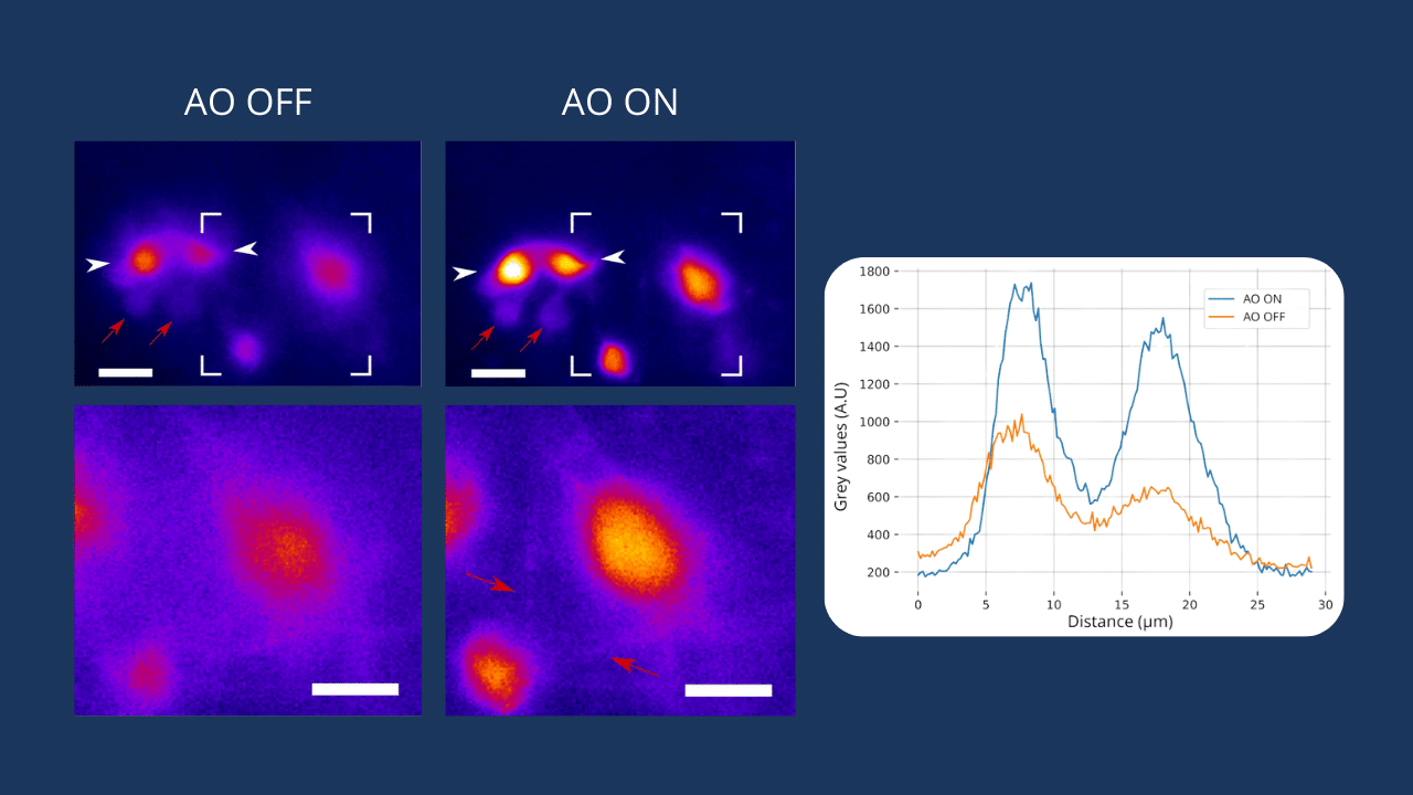
In this new study, an enhanced AO-LSFM set-up which does not require any guide star and is compatible with calcium neuroimaging is demonstrated.
Using dual labelling in the Drosophila brain, a strongly scattering sample :
– Structural and calcium signal images down to 80 and 40 μm were reported respectively, with an increased contrast of at least a factor of 2.
– Fast AO correction at a speed up to 10 Hz was demonstrated, positioning the approach as a game-changer in 3D imaging and dynamic process observation in depth in scattering samples.
Next steps and Future Frontiers
The instrumental approach designed by the team offers a compact and versatile prototype for integration into various LSFM setups. Ongoing testing on different configurations reveals the potential for a commercial AO add-on.
Researchers now delve into new challenges such as light-sheet thickness and excitation optimization, showcasing the continuous evolution of AO-LSFM technology for even greater image quality and sensitivity.
In conclusion, with its potential to unlock the mysteries of the brain, AO-LSFM is one of the technologies standing at the forefront of neuroimaging innovation. To address current & future challenges in this promising field, we are dedicated to developing tailored adaptive optics solutions to propel research forward.
You have questions regarding adaptive optics in microscopy? Do not hesitate to contact us!